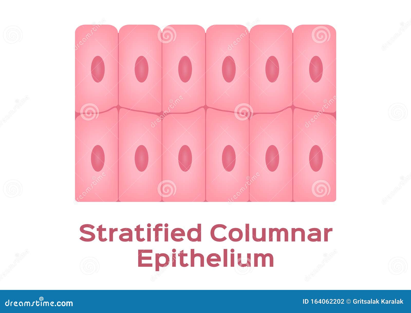
39 stratified columnar diagram
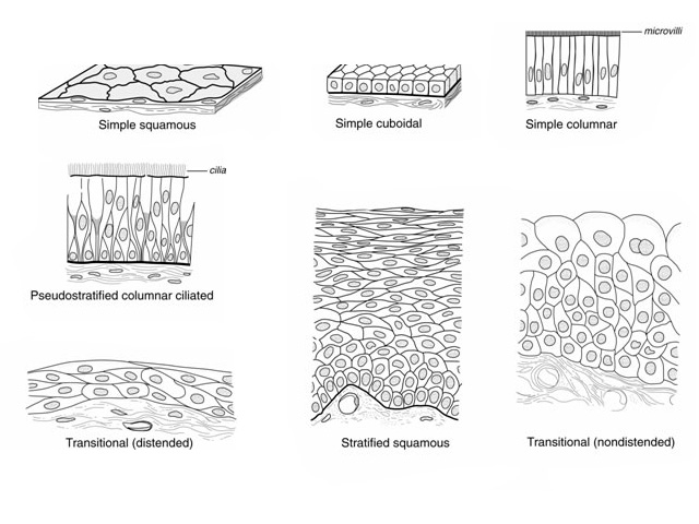
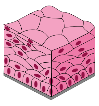
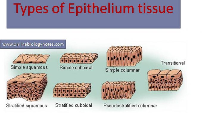
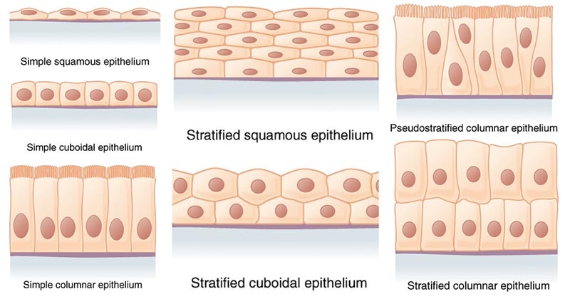
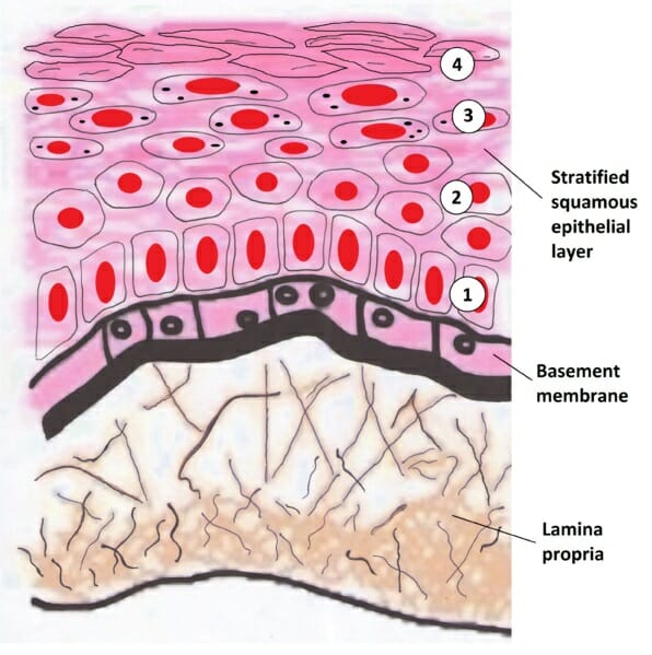
Stratified Squamous Epithelium Function, Structure ... The stratified squamous epithelium consists of cell layers in which the superficial layer consists of squamous epithelial cells while the underlying cell layers have various types of cells. The deepest layer is made up of columnar cells. This type of epithelial tissue covers body parts that are exposed to frequent frictional forces or stress. Stratified Columnar Epithelium Diagram | Quizlet Start studying Stratified Columnar Epithelium. Learn vocabulary, terms, and more with flashcards, games, and other study tools.
Tongue Histology - Connective Tissue and Taste Buds of ... 03.08.2021 · Tongue is a muscular organ of animal and important for prehension, mastication and deglutination of food. In the tongue histology, you will find the skeletal muscle that covered by mucosa membrane.Again, the mucosa membrane consists of stratified squamous epithelial lining which may be keratinized or non-keratinized in different parts.

Stratified columnar diagram
Simple Columnar Epithelium: A Labeled Diagram and ... The columnar epithelial cells are shaped like a column, with the height being greater than the width. They are also classified on the basis of the number of layers of cells. The tissues that comprise a single layer of cells are called simple, and the ones that have more than one layer are referred to as stratified. Oral: The Histology Guide This diagram shows across section through the lip and tooth, showing some of the main features of these structures. All of the oral mucosa is made up of a thick stratified squamous epithelium, supported by a lamina propria. The epithelium is thick because the epithelial lining of the oral cavity is subject to a lot of wear and tear. In mobile areas, such as the soft palate, underside of … Chapter 3 Diagram Practice Flashcards | Quizlet Ellie_Klumb. Chapter 8 Diagram Practice. 30 terms. Ellie_Klumb. Chapter 7 Vocabulary. 43 terms. Ellie_Klumb. Upgrade to remove ads. Only $35.99/year.
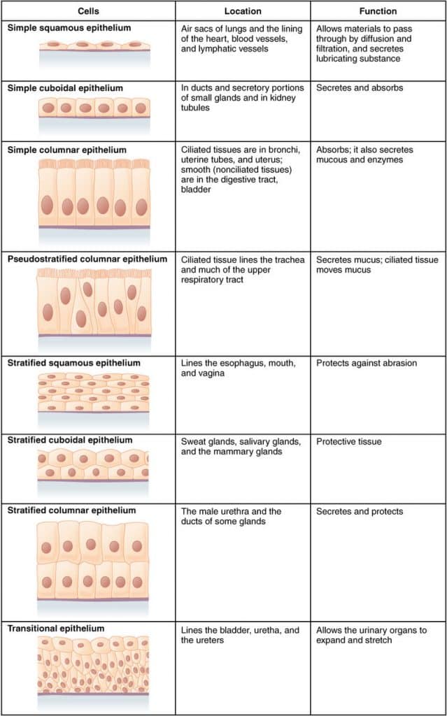
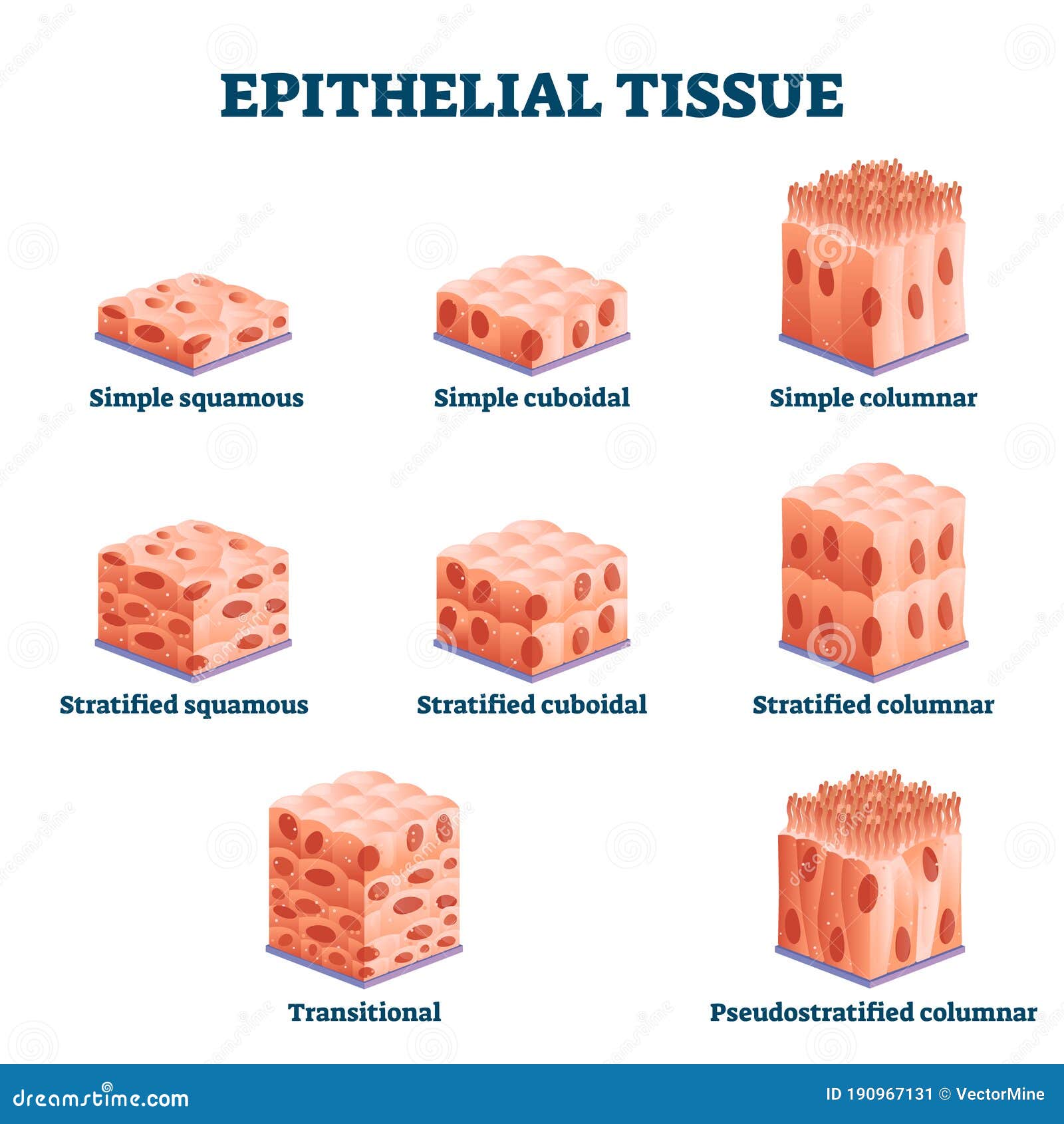
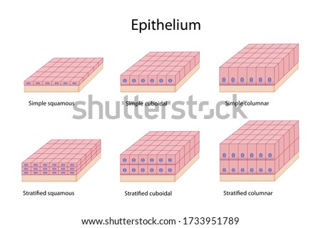
Stratified columnar diagram. Stratified Columnar Epithelium Diagram | Quizlet Start studying Stratified Columnar Epithelium. Learn vocabulary, terms, and more with flashcards, games, and other study tools. [Figure, Diagram of an acinus, as...] - StatPearls - NCBI ... Red cells covering acini and intercalated ducts are myoepithelial cells. Blue cells of intercalated duct represent single layer of cuboidal cells. Columnar cells of striated duct with basolateral membrane invaginations are represented by purple cells. Pseudo-stratified columnar cells of excretory duct are represented by green cells. Lab Quiz # 3 Diagram | Quizlet stratified columnar epithelium. provides abrasion protection of skin epidermis and oral cavity. stratified squamous epithelium. forms inner lining of urinary bladder and ureters. transitional epithelium. lines kidney tubules and ducts of salivary glands. simple cuboidal epithelium. Forms the lining of the stomach and small intestines . simple columnar epithelium. Two or three … Diagram To Show The Various Kinds Of Epithelium -- Simple ... Diagram To Show The Various Kinds Of Epithelium -- Simple Squamous, Stratified Squamous, Cuboidal, Columnar And Transitional Royalty Free Cliparts, Vectors, And Stock Illustration. Image 14742327. Text Filter Auto enhance Background removal Reset All Standard sizes S 857 x 559 px HIWEB scale to any size x scale to any size px M 2616 x 1705 px
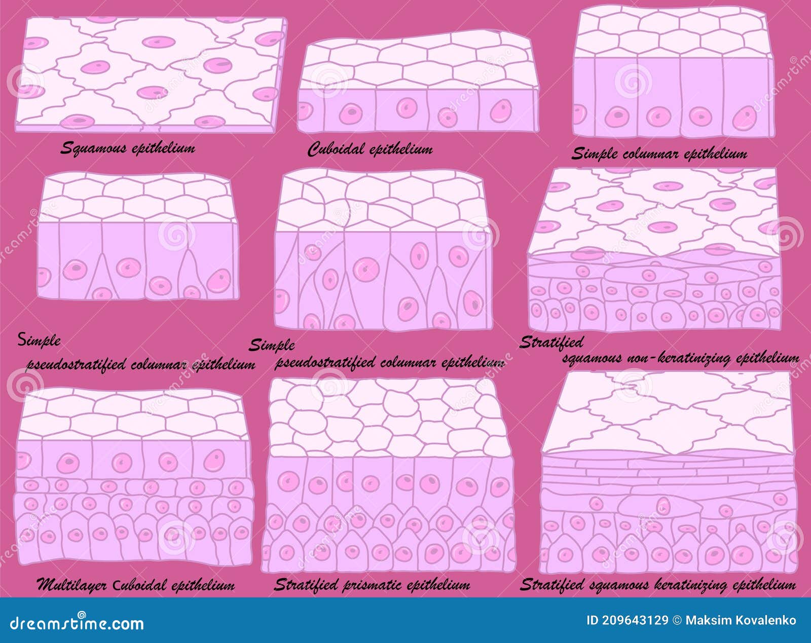
Exocrine gland - Wikipedia Exocrine glands are glands that secrete substances on to an epithelial surface by way of a duct. Examples of exocrine glands include sweat, salivary, mammary, ceruminous, lacrimal, sebaceous, prostate and mucous.Exocrine glands are one of two types of glands in the human body, the other being endocrine glands, which secrete their products directly into the … Stratified columnar epithelium- structure, functions, examples The stratified columnar epithelium consists of many layers of cells, where the cells in the deeper layers are irregular and of different shapes. In contrast, the cells in the apical layer are column-like in shape. The cells in the stratified columnar epithelium, as in the case of simple columnar epithelium are taller than they are wide. Stratified epithelium: Characteristics, function, types ... Stratified epithelium consists of two or more cell layers. There is a great amount of variability between the layers due to various cellular shapes and heights. The three types of cellular shapes are squamous, cuboidal, and columnar. Squamous cells have a width greater than the height and contain an ovoid, centered nucleus. Pseudostratified Columnar Epithelium under a Microscope ... Pseudostratified Columnar Epithelium under a Microscope with a Labeled Diagram 06/04/2022 by anatomylearner The pseudostratified columnar epithelium comprises a single layer of cells but seems to be multilayered. It is because different cellular heights and nuclei are also placed at a different levels.
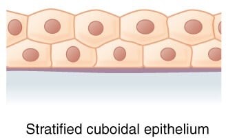
Simple Columnar Epithelium Labeled Diagram Squamous. stratified squamous diagram photo of endothelial cells. Squamous means scale-like. simple squamous. Bodytomy provides a labeled diagram to help you understand the structure and Simple Columnar Epithelium: Labeled Diagram and Function. Epithelium is a tissue that lines the internal surface of the body, as well as the internal organs. Histology Slides Identification from Different Organ ... The pseudostratified stratified columnar epithelium lines the ductus epididymis. You will find the smooth muscle fibers surrounding the ducts and sperms in the lumen of the duct. In the vas deference histology slide, you will find a stellate-shaped lumen and thick muscular coats. Development of Frog (With Diagram) | Vertebrates ... The neural plate cells change in shape and become elongated and arranged themselves into a columnar epithelium. The epidermal cells remain more or less flat and arranged as a stratified epithelium usually two cells thick. The edges of the neural plate become thickened and slightly raised above the general level as ridges called neural folds. The neural plate narrows … Stratified cuboidal epithelium- structure, functions, examples Stratified cuboidal epithelium has multiple layers of cells in which the apical layer is made up of cuboidal cells while the deeper layer can be either cuboidal or columnar. As in the case of stratified squamous epithelium , the cells in the deeper layers might be different than the layer on the top.
Merocrine - Wikipedia Merocrine (or eccrine) is a term used to classify exocrine glands and their secretions in the study of histology.A cell is classified as merocrine if the secretions of that cell are excreted via exocytosis from secretory cells into an epithelial-walled duct or ducts and then onto a bodily surface or into the lumen.. Merocrine is the most common manner of secretion.
Solved 1. Identify the type of epithelium indicated by the ... Stratified cuboidal 4. Identify the type of epithelium indicated by the diagram below: a. Simple columnar b. Stratified columnar c. Pseudostratified columnar d. Stratified cuboidal 50 um 5. Identify the structures incicated by the black arrows: a. Microvilli b. Cilia c. Villi d. None of the above 6.
Epithelial Tissue Structure, Types and Function (With ... III) STRATIFIED COLUMNAR EPITHELIUM Outermost layer is composed of pillar shaped cells, cells are nucleated. On the basis of presence of cilia this epithelium is of 2 types (1) Ciliated stratified columnar epithelium Eg. Buccopharyngeal cavity of Frog. Larynx (2) Non ciliated stratified columnar epithelium. Cilia absent on free end. Eg. Epiglottis
PDF WEEK 1: COVERING AND LINING EPITHELIA - University of New ... They are sketches from selected slides used in class from the teaching slide set. These labelled diagrams should closely follow the current Science courses in histology, anatomy and embryology and complement the virtual microscopy used in the current Medical course. © Dr Carol Lazer, April 2005 STEREOLOGY:SLICING A 3-D OBJECT SIMPLE TUBE
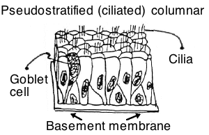
Pseudostratified Columnar Epithelium Histology - Jotscroll Pseudostratified columnar epithelium diagram Location of Pseudostratified Columnar Epithelium Ciliated Pseudostratified columnar epithelium lines the Bronchi Pseudostratified columnar epithelium is found in some parts of the auditory tube It is also found in the ductus deferens It also lines the membranous and penile parts of the male urethra
4.2 Epithelial Tissue - Anatomy & Physiology Stratified squamous epithelium is the most common type of stratified epithelium in the human body. The apical cells appear squamous, whereas the basal layer contains either columnar or cuboidal cells. The top layer may be covered with dead cells containing keratin. The skin is an example of a keratinized, stratified squamous epithelium.
Question 55 of the Science Practice Test for the TEAS The attached diagram shows four varieties of one type of tissue. Name the type of tissue and the four varieties. View. nervous: grey matter, white matter, dendritic, and ganglion . connective: adipose, cardiac, transitional, and bone. connective: adipose, blood, areolar, and bone. epithelial: transitional, stratified, columnar, and cuboidal. Previous Question Next Question Create a …
Stratified Columnar Epithelium Diagram | Quizlet Stratified Columnar Epithelium Diagram | Quizlet. Upgrade to remove ads. Only $35.99/year.
Solved Activity 3: Tissue Concept Map (#5-#21 ... - chegg.com For this one, follow the flow of the diagram to fill in the correct responses to organize the tissue information. START with the word TISSUE in the center. Question: Activity 3: Tissue Concept Map (#5-#21) The concept map is on the next page with space to write the answers. Concept maps are a way to organize your information and everyone does ...
Tonsil Histology Slide with Labeled Diagram - Histological ... The stratified squamous consists of many layers of cells (columnar, polyhedral, and squamous). There are flat cells (squamous) with elliptical nuclei present in the superficial layer of the stratified squamous. The pseudostratified columnar epithelium consists of cells of different shapes and heights lying on the basement membrane.
Epithelial Tissue: Structure with Diagram, Function, Types ... Compound (Stratified) Epithelium- it is made up of two or more than two layers of cells and mostly has a protective function. The glandular epithelium is made up of cuboidal or columnar cells. They are specialised for secretion. Unicellular- isolated glandular cells, e.g. goblet cells Multicellular- a cluster of cells, e.g. salivary glands
Simple Squamous Epithelium under a Microscope with a ... From the lung parenchyma labeled diagram, you might identify the following structures - Simple squamous epithelium lining of the lung alveoli (within the parenchyma), A connective tissue basement membrane beneath the simple squamous epithelium lining, The lumen of the lung alveoli, and The cytoplasm of the simple squamous epithelium cells.
Solved A B C D E Reference: Ref 4-2 In the diagram shown ... Simple cuboidal Stratified squamous Transitional Simple squamous Stratified columnar This problem has been solved! See the answer See the answer See the answer done loading
Pseudostratified Columnar Epithelium Function & Location ... Pseudo-Stratified Columnar Epithelium. The epithelial tissue made up of a single layer of epithelial cells of different heights is known as the pseudostratified columnar epithelium.The position of ...
Ciliated Epithelium - Concept, Structure, Function and ... Pseudostratified ciliated columnar epithelia are the tissues that are formed by only one layer of cells and give the appearance of being made from multiple layers, especially when seen in a cross-section. The nuclei of the epithelial cells are at very different levels that will lead to the illusion of being stratified.
Pseudostratified Columnar Epithelium - AnatomyZone Pseudostratified Columnar Epithelium Articles / By Dr Peter de Souza This diagram represents pseudostratified columnar epithelium . This is a special type of simple epithelium called pseudostratified epithelium as it resembles stratified epithelium due to the positioning of the cellular nuclei, but is comprised of only a single layer of cells.
PseudoStratified columnar epithelium | Psychology notes ... Pseudostratified epithelium is also sometimes referred to as respiratory epithelium, since ciliated pseudostratified columnar epithelia is mainly found in the larger respiratory airways of the nasal cavity, trachea and bronchi. Find this Pin and more on File Diagrams by Histopedia. Histology Slides. Psychology Notes. Nasal Cavity. Anatomy. Larger.
Chapter 3 Diagram Practice Flashcards | Quizlet Ellie_Klumb. Chapter 8 Diagram Practice. 30 terms. Ellie_Klumb. Chapter 7 Vocabulary. 43 terms. Ellie_Klumb. Upgrade to remove ads. Only $35.99/year.
Oral: The Histology Guide This diagram shows across section through the lip and tooth, showing some of the main features of these structures. All of the oral mucosa is made up of a thick stratified squamous epithelium, supported by a lamina propria. The epithelium is thick because the epithelial lining of the oral cavity is subject to a lot of wear and tear. In mobile areas, such as the soft palate, underside of …
Simple Columnar Epithelium: A Labeled Diagram and ... The columnar epithelial cells are shaped like a column, with the height being greater than the width. They are also classified on the basis of the number of layers of cells. The tissues that comprise a single layer of cells are called simple, and the ones that have more than one layer are referred to as stratified.
















Comments
Post a Comment