43 areolar tissue diagram
Areolar Tissue: Definition, Functions, Structure & Location. Cells are the structural and functional units that cover the entire human body. Instead of the cells having an individual structure, they are grouped into clusters. We call these clusters 'tissues'. Biologically, tissues are groups of cells that have a collective function. Download scientific diagram | A graft of perifascial areolar tissue (PAT). (A) A layer of loose connective tissue was elevated from the surface of the femoral fascia. (b) Macrographic and (C ...
Draw a labelled diagram of areolar connective tissue. structural organisation in animals; class-11; Share It On Facebook Twitter Email. 1 Answer +1 vote . answered Feb 5, 2020 by KumariJuly (53.6k points) selected Feb 5, 2020 by Ritik01 . Best answer. Diagram of areolar connective tissue ...

Areolar tissue diagram
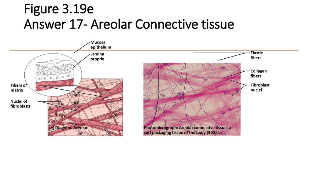
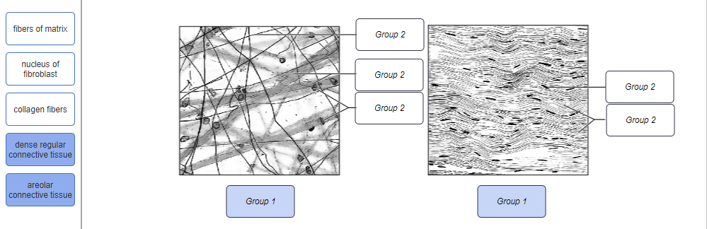
Figure 3.19e Connective tissues and their common body locations. Mucosa epithelium Lamina propria Fibers of matrix Nuclei of fibroblasts Elastic fibers Collagen fibers Fibroblast nuclei (e) Diagram: Areolar Photomicrograph: Areolar connective tissue, a soft packaging tissue of the body (270×) Areolar connective tissue is the most familiar type of connective tissue in vertebrates. It carries organs in place and attaches epithelial tissue to other underlying tissues. For example, it creates telae, such as the tela submucosa and tela subserosa, which attach mucous and serous membranes to the muscular layer. (areolar tissue is found in both the dermis and sub-cutaneous layers of the skin - see diagram). In this lesson, you'll explore what areolar connective tissue is, what elements Simple Cuboidal Epithelium: Location, Structure & Function.the first subclass of connective tissue proper is loose connective tissue.
Areolar tissue diagram. Please Give A Detailed Diagram Of Areolar And Adipose Connective Tissue With Proper Explanation Of Each Component Of It Biology Topperlearning Com Zh17wp88. With Help Of Neat Labelled Diagram Describe The Structure Of Areolar Connective Tissue Brainly In. 33 2b Connective Tissues Loose Fibrous And Cartilage Biology Libretexts. Areolar tissue is found in many locations around the body. One important area is the skin (areolar tissue is found in both the dermis and sub-cutaneous layers of the skin - see diagram).The areolar tissue located in the skin binds the outer layers of the skin to the muscles beneath. Areolar connective tissue. wraps: blood vessels nerves glands ... As stated earlier, the areolar tissue is the most widely distributed connective tissue in the body. It is present under the skin as subcutaneous tissue in between and around muscles, nerves and blood vessels in sub-mucosa of gastrointestinal tract and respiratory tract, in the bone marrow, between the lobes and lobules of compound glands and in ... How to draw a Diagram of Areolar Tissue in exam is the topic. This is the well labelled diagram of structure of Areolar Tissue. This is a well labelled diagr...
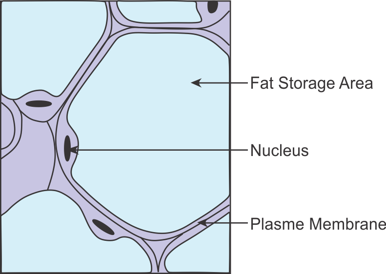
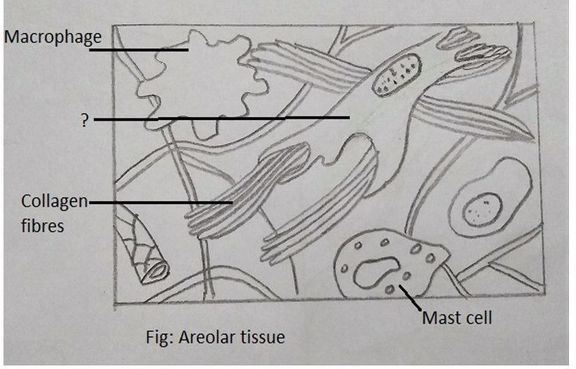
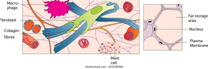
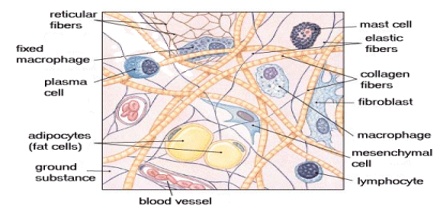
Areolar connective tissue serves quite a few functions and, because of this, is found in various places of the body, such as surrounding your blood vessels, nerve bundles, muscles, and organs. It ... areolar tissue diagram - Google Search. Denver Jaque. Debut Invitation. Invitation Cards. Tissue Biology. Human Tissue. Tissue Types. Drawing Sketches. Drawings. Body Tissues ... cell and tissue studies, and an overview of all the body systems. Intended as a survey course for certain allied health and social service programs, and as a general ... Helps in the repair of tissues. Adipose tissue Found beneath the skin, around the kidneys and other internal organs such as intestines. Cells are filled with fat globules situated in a large central vacuole of a cell, pushing the cytoplasm and nucleus to the periphery. A loose framework of areolar tissue supports these cells. Areolar Tissue. Areolar tissue is the most widely distributed solid connective tissue in the body. Which makes sense because areolar tissue is present under the entire skin. And areolar tissue also supports the epithelium. Areolar tissue contains randomly spread out fibers, fibroblasts, mast cells, and macrophages.
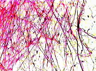
Learn areolar tissue with free interactive flashcards. Choose from 88 different sets of areolar tissue flashcards on Quizlet. areolar connective tissue, adipose connective tissue, reticular connective tissue, dense irregular connective tissue, dense regular connective tissue, elastic connective tissue Name the tissues that are listed under connective tissue proper The given diagram is the connective tissue - areolar tissue.Areolar connective tissue is found between the skin and muscles, around blood vessels and nerves and in the bone marrow. It fills the space inside the organs, supports internal organs and helps in repair of tissues. Areolar connective tissue is a loosely arranged connective tissue that is widely distributed in the Body and contains collagen fibres, reticular fibres and a few elastic fibres embedded in a thin, almost fluid-like ground substance.
Suffix Mds Dangerous Area Of Scalp 4th Layer Loose Areolar Tissue Because It Gives Passage To Emissary Veins Which Course Here May Transmit Infection From Scalp To Intracranial Venous Sinuses Facebook
Connective Tissue Diagrams by Julie Ridge are part of my human anatomy series. This set contains the following 10 diagrams: Adipose tissue, indicating adipocytes; Areolar tissue, indicating fibrocytes, collagen fibers, mast cell and elastic fibers; Blood, indicating red blood cells, white blood cells, platelets and plasma; Dense Irregular Connective Tissue, indicating collagen fiber bundles ...

Areolar Tissue Tissue Biology Human Anatomy And Physiology Anatomy And Physiology
Areolar connective tissues hold organs in place and attaches epithelial tissue to other underlying tissues. It also serves as a reservoir of. In this lesson, you'll explore what areolar connective tissue is, what elements Simple Cuboidal Epithelium: Location, Structure & Function.Dec 11, · Diagram Of Areolar Tissue.
Easy Connective
Areolar Connective Tissue Definition. Areolar connective tissue is a loosely arranged connective tissue that is widely distributed in the Body and contains collagen fibers, reticular fibers and a few elastic fibers embedded in a thin, almost fluid-like ground substance. The areolar connective tissue is a subtype of loose connective tissue.

15 Anatomy Ideas Anatomy Loose Connective Tissue Collagen Fibers
Human Anatomy Connective Tissue Diagrams By Julie Ridge Designs Teachers Pay Teachers Human Anatomy Anatomy Adipose Tissue. Areolar Tissue Diagram Google Search Tissue Tissue Types Collagen Fibers. Simple Cuboidal Sldie Labeled Histology Epithelial Tissues Histology Slides Study Of Tissues Medicine Notes. Integumentary System Diagram Google ...

Areolar Connective Tissue Model Diagram Quizlet
07.09.2021 · Ciliated epithelium is an important tissue found in various parts of the body and aids in everyday health. Explore what ciliated epithelial is and its function, its structure using a diagram, why ...
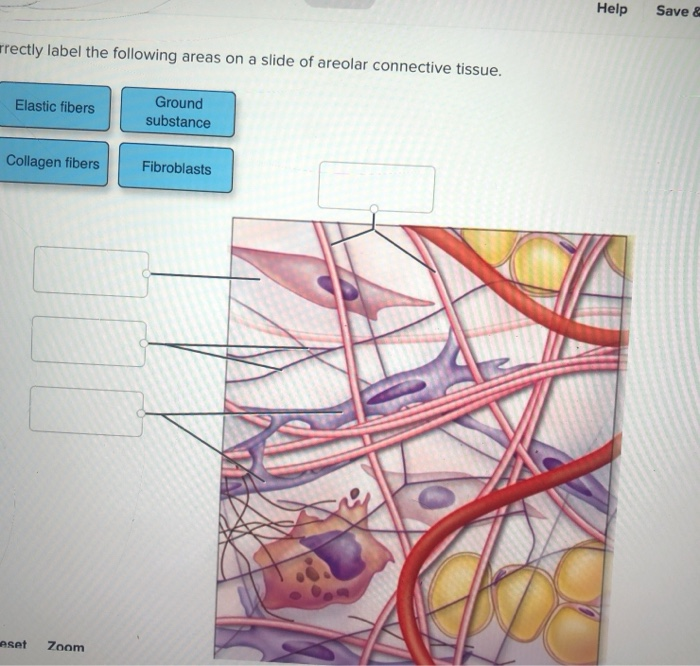
Solved Ectly Label The Following Areas On A Slide Of Areolar Chegg Com
Areolar Connective Tissue Diagram. This diagram shows areolar connective tissue serving as a medium between epithelial tissue and blood vessels. Cellular components (such as fibroblasts) and the ...

Section Of Septum Transversum Showing Abundant Loose Areolar Tissue L Download Scientific Diagram
Areolar Tissue Diagram. Areolar Connective Tissue Definition Areolar connective tissue is a loosely arranged. To bind parts together is the primary function of areolar tissue. Other functions are to provide strength, elasticity, support to the parts where this tissue is present. Figure Diagram of areolar tissue as it appears in microscopic section.
Please Give A Detailed Diagram Of Areolar And Adipose Connective Tissue With Proper Explanation Of Each Component Of It Biology Topperlearning Com Zh17wp88
Areolar tissues are widely distributed in the body and primarily function as a packing material between other tissues. Functions of the Areolar Connective Tissue. The areolar connective tissue is a type of connective tissue that is present throughout the human body. It provides support and helps to protect organs, muscles, and many other tissues.

Connective Tissue Zoology
27.06.2012 · Figure 1 Diagram of the ductal system. Multiple ductal systems drain into a ductal sinus within a segment and subsequently empty into the nipple. The breast is supported by Cooper ligaments, which run from the chest wall toward the nipple in a radial fashion. The pectoralis major muscle represents the most posterior boundary of the breast. TDLU = terminal ductal–lobular unit. Figure 1 ...

Draw A Labeled Diagram Of Areolar Tissue Brainly In
The areolar tissue is found beneath the dermis layer and is also underneath the epithelial tissue of all the body systems that have external openings. It is also a component of the lamina propria of the digestive and respiratory tracts, the mucous membranes of reproductive and urinary system, the stroma of glands, and the hypodermis of the skin.. It is also found in the mesentery which is ...

Areolar Connective Tissue Diagram Quizlet
Areolar Tissue: Areolar tissue is the most widely distributed connective tissue in the animal body. It is present under the skin and supports the epithelium. It contains randomly distributed fibres, fibroblasts, mast cells and macrophages. It supports the organs present in the abdominal cavity, fills the space between muscle fibres and wraps ...

Scalp Radiology Reference Article Radiopaedia Org
ADVERTISEMENTS: The following points highlight the ten main varieties of connective tissues of human body. They are: 1. Areolar Tissue 2. Adipose Tissue 3. White Fibrous Tissue 4. Yellow Elastic Tissue 5. Reticular Tissue 6. Blood and Haemopoietic Tissue 7. Cartilage 8. Jelly-Like Connective Tissue 9. Osseous Tissue or Bone 10. Reticulo-Endothelial Tissue. 1. Areolar […]

Areolar Tissue Diagram Google Search Tissue Tissue Types Collagen Fibers
Breast anatomy is complex and intricate, involving various types of tissue, including milk ducts, lymph vessels, the nipple and areola, and other structures. Here's what to know about the many internal and external parts of the breast, their purpose, and the medical conditions that can affect them.
Zoology Exam Questions And Answers Sanfoundry
(areolar tissue is found in both the dermis and sub-cutaneous layers of the skin - see diagram). In this lesson, you'll explore what areolar connective tissue is, what elements Simple Cuboidal Epithelium: Location, Structure & Function.the first subclass of connective tissue proper is loose connective tissue.
Draw A Labelled Diagram Of Areolar Connective Tissue Sarthaks Econnect Largest Online Education Community
Areolar connective tissue is the most familiar type of connective tissue in vertebrates. It carries organs in place and attaches epithelial tissue to other underlying tissues. For example, it creates telae, such as the tela submucosa and tela subserosa, which attach mucous and serous membranes to the muscular layer.

Areolar Connective Tissue Diagram Quizlet
Figure 3.19e Connective tissues and their common body locations. Mucosa epithelium Lamina propria Fibers of matrix Nuclei of fibroblasts Elastic fibers Collagen fibers Fibroblast nuclei (e) Diagram: Areolar Photomicrograph: Areolar connective tissue, a soft packaging tissue of the body (270×)
Connective Tissue Diagram Images Stock Photos Vectors Shutterstock

A Graft Of Perifascial Areolar Tissue Pat A A Layer Of Loose Download Scientific Diagram

Fibrous Connective Tissue

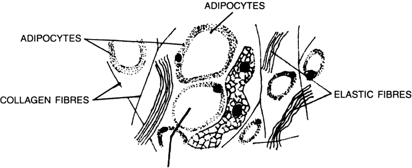
Difference Between Areolar And Adipose Tissue Compare The Difference Between Similar Terms

00001942 Jpg
Areolar Connective Tissue

How To Draw Areolar Tissue How To Draw Areolar Connective Tissue Draw Diagram Of Areolar Tissue Youtube

Classification Of Connective Tissue Physiotherapy Milan

Areolar Connective Tissue Function Location What Is Areolar Connective Tissue Video Lesson Transcript Study Com

Diagram Of Adipose And Areolar Tissue Brainly In
Tissue Diagram Quiz Review Ppt Download
A Diagram Of Areolar Connective Tissue Is Given Below Img Src Https D10lpgp6xz60nq Cloudfront Net Physics Images Nta Neet Set 35 E03 006 Q01 Png Width 80 Identify The Option With Correct Labeling
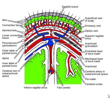
Scalp Anatomy Structure Nerve Supply Arterial Supply
Solved Drag The Labels Onto The Diagram To Identify The Chegg Com
What Are Areolar Tissue And Adipose Tissue Where They Are Located
Loose Connective Tissue Assignment Point

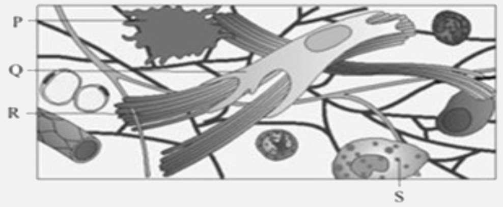
Parts A B C And D Of The Areolar Connective Tissue Are Shown In The Diagram Selection The Option Which Gives Correct Identification Along With Its Functions Characteristics

Connective Tissue Anatomy And Physiology

Loose Areolar Ct Diagram Diagram Quizlet
1
Diseases Of The Ovaries Their Diagnosis And Treatment A Umbilicus H Skin C Linea Alba D Symphysis C Peritoneum Superficial Layer Of Areolar Tissue G Deep Layer Of Ditto H Areolar Tissue

Areolar Connective Tissue Diagram Quizlet

Schematic Drawing Of Perifascial Areolar Tissue Pat Download Scientific Diagram
How Do Areolar Tissue Qualify As Connective Tissues Quora

A Diagram Of Areolar Connective Tissue Is Given Below Img Src Https D10lpgp6xz60nq Cloudfront Net Physics Images Nta Neet Set 35 E03 006 Q01 Png Width 80 Identify The Option With Correct Labeling

With Help Of Neat Labelled Diagram Describes The Structure Of Areolar Connective Tissue Brainly In

Areolar Connective Tissue


Comments
Post a Comment